


Dental Anatomy: 101
Learn more about your teeth!

What Are the Parts of a Tooth?
Check out the definitions of the anatomical terms in the diagram above.
Enamel - Dental enamel is the hard thin translucent layer that serves as protection for the dentin of a tooth. It is made up of calcium salts. It is the hardest substance in the body.
Dentin - Dentin is the hard, dense, calcium like material that makes up most of the tooth underneath the enamel and cementum. It is harder and denser than bone. It is the second layer of the tooth.
Anatomical Crown - The natural, top part of a tooth, which is covered in enamel and is the part that you can see extending above the gum line.
Pulp Chamber - The area within the natural crown of the tooth where the tooth pulp resides. It contains blood vessels and nerve tissue.
Gingiva - Also known as gums - the soft tissues that cover part of the tooth and bone. Gingiva helps protect the teeth from any infection or damage from food and everyday interactions with the outer world.
Neck - The area of the tooth where the crown joins the root.
Root Canal - Not to be confused with Root Canal Treatment, the root canal is a space inside your tooth root that is filled with nerves, blood vessels, and soft tissue.
Jawbone (Alveolar Bone) - The part of the jaw that surrounds the roots of the teeth and which is connected to the tooth by the periodontal ligament.
Cementum - A thin layer of hard connective tissue that covers the outer surface of the root. This substance is yellow and not as hard as enamel or dentin.
Periodontal Ligament - - A system of fibrous connective tissue between the cementum and bony socket.
The Anatomy of a Tooth
Your teeth are composed of hard (calcified) and soft (non-calcified) dental tissues. Enamel, dentin and cementum are hard tissues. Pulp, or the center of the tooth that contains nerves and blood vessels ?is a soft tissue. Each part of the tooth plays a vital role, whether it is to chew or tear food, speak, or even protect the tooth itself from decay.
Tooth Charts
Our mouths contain teeth of various shapes, sizes, and locations in the jaw. Each type of tooth is designed to perform different functions, like biting, tearing, and chewing. How teeth are shaped and aligned affect your smile, speech, and facial shape. People are usually born with 20 baby (primary) teeth, which start to erupt around about 6 months of age and shed at different times throughout childhood. By age 21, all 32 of the permanent teeth (including wisdom teeth) have usually erupted.
Primary (Baby) Teeth
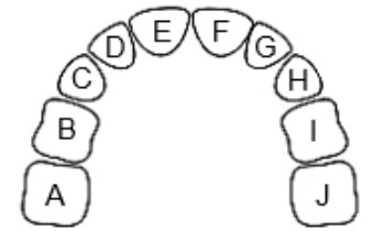

| Number | Erupt | Shed | Upper Teeth |
|---|---|---|---|
| A,J | 25-33 mos | 10-12 yrs | Second Molar |
| B,I | 13-19 mos | 9-12 yrs | First Molar |
| C,H | 16-22 mos | 10-12 yrs | Canine (cuspid) |
| D,G | 9-13 mos | 7-8 yrs | Lateral Incisor |
| E,F | 8-12 mos | 6-7 yrs | Central Incisor |
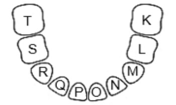

| Number | Erupt | Shed | Lower Teeth |
|---|---|---|---|
| K,T | 23-31 mos | 10-12 yrs | Second Molar |
| L,S | 14-18 mos | 9-11 yrs | First Molar |
| M,R | 17-23 mos | 9-12 yrs | Canine (cuspid) |
| N,Q | 10-16 mos | 7-8 yrs | Lateral Incisor |
| O,P | 6-10 mos | 6-7 yrs | Central Incisor |
Permanent (Adult) Teeth
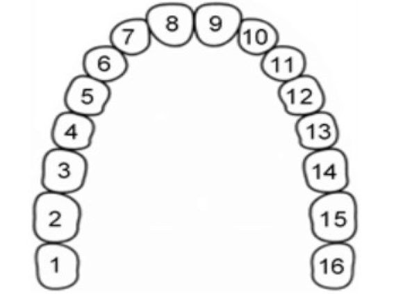
| Number | Erupt | Upper Teeth |
|---|---|---|
| 1,16 | 17-21 yrs | Third Molar (Wisdom Teeth) |
| 2,15 | 12-13 yrs | Second Molar (12 yr molar) |
| 3,14 | 6-7 yrs | First Molar (6 yr molar) |
| 4,13 | 10-12 yrs | Second Premolar |
| 5,12 | 10-11 yrs | First Premolar |
| 6,11 | 11-12 yrs | Canine (cuspid) |
| 7,10 | 8-9 yrs | Lateral Incisor |
| 8,9 | 7-8 yrs | Central Incisor |
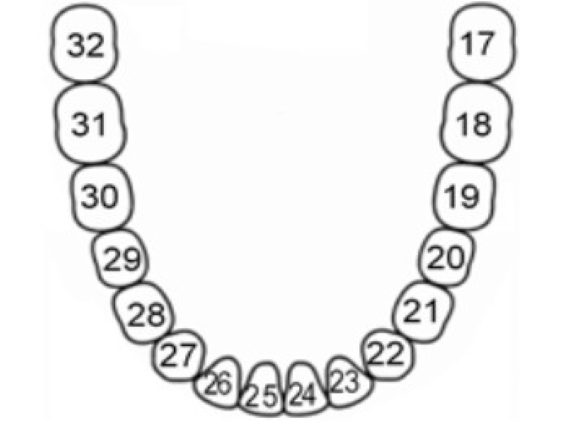
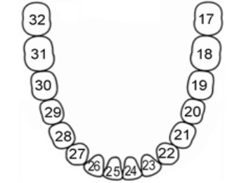
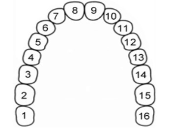
| Number | Erupt | Lower Teeth |
|---|---|---|
| 17,32 | 17-21 yrs | Third Molar (Wisdom Teeth) |
| 18,31 | 12-13 yrs | Second Molar (12 yr molar) |
| 19,30 | 6-7 yrs | First Molar (6 yr molar) |
| 20,29 | 10-12 yrs | Second Premolar |
| 21,28 | 10-11 yrs | First Premolar |
| 22,27 | 11-12 yrs | Canine (cuspid) |
| 23,26 | 8-9 yrs | Lateral Incisor |
| 24,25 | 7-8 yrs | Central Incisor |
Note: The information in this document is not meant to replace the advice of your dentist or another licensed healthcare professional. Talk to your dentist for any specific dental advice.